Friday 20 September 2013

Ionotropic Glutamate Receptors

Glutamate is the major excitatory neurotransmitter in vertebrate brains. Ionotropic glutamate receptors (iGluRs) are the ligand-gated ion channels that mediate the majority of fast synaptic transmission. AMPARs and NMDARs are named after the drugs (agonists) that were used to discover them. They are well known as regulators of synaptic plasticity, but I am not sure how such regulation is implemented. For example, NMDARs may be on both sides of a synaptic cleft, i.e. on the bouton and spine. Short term plasticity is mostly due to changes to the bouton. Long term plasticity is mostly due to changes in the spine. The precise location, quantity and quality of receptors (their subunit composition and/or phosphorylation) ... and structural changes, e.g. in spine morphology, may contribute to the strength of the synapse.

Key point: ionotropic receptors are gated by a neurotransmitter, not by voltage. Although some NMDARs are subject to a Mg2+ block, they are voltage dependent but not voltage-gated per se ... the channel itself does not open/close due to depolarisation, none of the NMDAR subunits include voltage sensor, instead the Mg2+ ion falls out into extracellular space. In contrast, a voltage-gated channel changes its conformation (opens/closes) due to a change in voltage (Vm).

Note: glial NMDARs have a weak Mg2+ block at physiological concentrations; their channels can be open at resting membrane potential (Karadottir et al., 2005; Lalo et al., 2006).

Receptor family NC-IUPHAR subunit nomenclature:

  • AMPA: GluA1 to GluA4
  • Kainate: GluK1 to GluK5
  • NMDA: GluN1, GluN2A, GluN2B, GluN2C, GluN2D, GluN3A, GluN3B

The NMDA receptor forms a heterotetramer between two GluN1 and two GluN2 subunits, two obligatory GluN1 subunits and two regionally localised GluN2 or GluN3 subunits (in most cases). However, NMDARs may have 4 or 5 subunits. There are eight variants of the GluN1 subunit and four variants of GluN2 subunit.

GluN2 subunits are expressed differentially across various cell types and control the electrophysiological properties of the NMDA receptor. One particular subunit, GluN2B, is mainly present in immature neurons and in extrasynaptic locations; it contains the binding-site for the selective inhibitor ifenprodil.

However, AMPARs and NMDARs can be built from various combinations of subunits.

The subunit composition of iGluRs and their density at synapses are major determinants of fast synaptic transmission and synaptic plasticity. This is because the subunit stoichiometry determines channel function, trafficking to synapses, and synapse-specific receptor expression. According to recent research RNA editing of AMPA-type iGluRs plays a central role for in these processes, which is currently under investigation.


Karadottir R, Cavelier P, Bergersen LH, Attwell D (2005). NMDA receptors are expressed in oligodendrocytes and activated in ischaemia. Nature 438: 1162–1166.

Lalo U, Pankratov Y, Kirchhoff F, North RA, Verkhratsky A (2006). NMDA receptors mediate neuron-to-glia signaling in mouse cortical astrocytes. J Neurosci 26: 2673–2683.
Presynaptic NMDA Receptors

NMDARs can be found on dendritic spines and many other locations. Postsynaptic locations are logical given that presynaptic neurons release glutamate. However, they can also be found on or near presynaptic terminals (boutons). In this case they can act as "autoreceptors" that provide direct feedback to the presynaptic terminal and, possibly, indirect feedback to the soma.

In a paper by McGuinness et al, 2010, the authors reported that preNMDARs allow more Ca2+ to enter the presynaptic terminal, elevate [Ca2+]i, and increase the probability of vesicle release during an action potential.

McGuinness, L, Taylor, C, Taylor, RDT, Yau, C, Langenhan, T, Hart, ML, Christian, H, Tynan, PW, Donnelly, P, and Emptage, NJ (2010). Presynaptic NMDARs in the hippocampus facilitate transmitter release at theta frequency. Neuron, 68(6):1109-27.

Saturday 14 September 2013

AMPA Receptors

Note: ligand-gated ionotropic receptors are classified into three superfamilies: cys-loop receptors, ionotropic glutamate receptors, and ATP-gated channels.

Other receptors are metabotropic receptors (which depend on ligands, but use second messengers), voltage-gated ion channels (which depend on membrane potential), and stretch-activated ion channels (which depend on mechanical deformation of the cell membrane).

AMPARs are ionotropic glutamate-gated receptors (iGluRs); they are named after the selective agonist alpha-amino-3-hydroxy-5-methyl-4-isoxazolepropionic acid (AMPA).

These receptors mediate fast synaptic transmission in the central nervous system. They are non-selective cation channels, i.e. they allow the passage of Na+ and K+. The influx of Na+ and efflux of K+ via this channel yields an equilibrium potential near 0 mV.

Each AMPAR has four sites to which an agonist (such as glutamate) can bind, one for each subunit. The channel opens when two sites are occupied, current increases as three and four sites are occupied. After the channel opens it may undergo rapid desensitisation, which stops the current.

"The AMPAR's permeability to calcium and other cations, such as sodium and potassium, is governed by the GluR2 subunit. If an AMPAR lacks a GluR2 subunit, then it will be permeable to sodium, potassium, and calcium. The presence of a GluR2 subunit will almost always render the channel impermeable to calcium. This is determined by post-transcriptional modification - RNA editing - of the Q/R editing site of the GluR2 mRNA" [wikipedia].

The prevention of calcium entry into the cell on activation of GluR2-containing AMPARs is thought to guard against excitotoxicity.

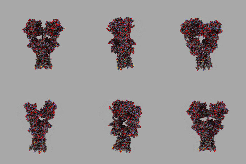
The AMPA receptor GluA2 (GluR2) tetramer was the first and, as of 15 September 2013, only glutamate receptor ion channel to be crystallised.

Image: wikipedia

Tuesday 10 September 2013

Unconventional neurotransmitters and retrograde signalling

Most neurotransmitters are released from a presynaptic terminal via "exocytosis". They diffuse across extracellular space and, typically, bind to postsynaptic receptors. However, not all neurotransmitters are presynaptic.

Anandamide and 2-AG are endocannabinoids. They inhibit LTP (which impairs memory, for example). Endocannabinoid production can be stimulated by a rise in [Ca2+] in postsynaptic neurons. Endocanninoid release is not well understood, but after release from a postsynaptic terminal these transmitters can bind to cannabinoid receptors such as CB1 receptors (a GPCR) on the presynaptic neuron, or other nearby cells.

Also, nitric oxide (NO) can permeate the plasma membrane of a postsynaptic neuron and diffuse into nearby cells, including the presynaptic terminal. NO acts intracellularly, so it is often considered to be "second messenger" rather than a neurotransmitter.

Hippocampus

The hippocampus has been used for many years to study synaptic plasticity. It is well known as being critical for memory. If the hippocampus is damaged, new declarative memories cannot formed. But it also critical for spatial learning, awareness, and navigation. For example, the rat hippocampus has been used to "bio-inspire" the RatSLAM software for robots: Milford, Robot Navigation from Nature Simultaneous Localisation, Mapping, and Path Planning Based on Hippocampal Models, 2008.

This structure is popular. It is "experimentally accessible" (to some techniques), and it is well suited to the study of synaptic plasticity due to its laminar organisation, the circuitry (connectivity between neurons) remains intact when brain slices are made.

Its cross-section has a very defined laminar structure; layers are visible where rows of pyramidal cells are arranged. In general, the connections within the hippocampus are uni-directional. They form well-characterised closed loops that start in the adjacent entorhinal cortex.

The different cell layers and sections are defined connections. The main pyramidal cell layers are the CA1-4 regions (principally CA1 and CA3), and the dentate gyrus.

Diagram below represents a slice of rodent hippocampus. It shows the major regions, excitatory pathways, and synaptic connections. Long-term potentiation has been observed at each of the three synaptic connections shown here.

Image: Purves et al, 2001.

Do all synaptic vesicles contain ATP?

"Interestingly, all synaptic vesicles contain ATP, which is co-released with one or more “classical” neurotransmitters. This observation raises the possibility that ATP acts as a co-transmitter. It has been known since the 1920s that the extracellular application of ATP (or its breakdown products AMP and adenosine) to neurons can elicit electrical responses" [Purves et al, Neuroscience, 2nd Edition, 2001]. The statement "all synaptic vesicles contain ATP" may sound dubious, and a professor who studies P2X receptors said "not true", but page 131 of 5th Edition (2012) contains the same text. This does not sound like a typographical error.

Glutamate is the most common excitatory neurotransmitter in the CNS and, by definition, it is stored in presynaptic vesicles. This raises an obvious question. Suppose that ATP is stored in all vesicles. They why is ATP stored with glutamate? And why would it be stored in all vesicles?

ATP binds to P2X ionotropic and P2Y metabotropic receptors. Adenosine binds to P1 receptors. Four subtypes of P1 receptors have been cloned: A1, A2A, A2B, and A3. Receptors for ATP and adenosine are widely distributed in the nervous system and other tissues (Burnstock and Sawynok 2010).

Caffeine and theophylline block adenosine receptors, so you might guess that adenosine makes you sleepy.

The other two classes of purinergic receptors are GPCRs; adenosine is the agonist for adenosine receptors, e.g. A1 and A2A, and ATP is the agonist for the the P2Y receptors.

If it is true that all synaptic vesicles contain ATP, then there must be a reason.


Burnstock and Sawynok 2010, Adenosine Triphosphate and Adenosine Receptors in Pain, Pharmacology of Pain, IASP Press, Seattle, p303.

P2X Receptors

The main points: The P2X receptor is ionotropic. When ATP binds to a P2X receptor, Ca2+ rushes into the cell.

The structure of P2X receptors (ionotropic) is unusually simple. Each subunit has only two transmembrane domains, and only three of these subunits are required to form a trimeric receptor.

[Image: wikipedia]

Seven separate genes coding for P2X subunits, named P2X1 through P2X7, have been identified. Only some combinations of three subunits are used to build this trimeric receptor, e.g. P2X7 homomeric, and P2X1/5 heteromeric receptors. Functional P2X6 homomeric and P2X7 heteromeric receptor cannot be formed.

Although the P2X pore forms a nonselective cation channel, it is especially permeable to calcium.

This receptor mediates excitatory postsynaptic responses.

They are widely distributed in CNS and PNS neurons.

P2X receptors are also present on astrocytes.

They may play a role in mechanosensation and pain in sensory nerves, but their function in most other cells is not known.

Neurotransmitters

Glutamate is the major excitatory neurotransmitter in the brain.

GABA and glycine are the major inhibitory transmitters in the brain.

For the moment, just consider the amino acids in the image below. Glutamate and aspartate are very similar, glycine too. If aspartate or glutamate levels are too high, then some neurons will be excited to death in a process known as "excitotoxicity".

Glutamate is an agonist for NMDA receptors.

Health note: aspartame is the chemical name for the brand names of artificial sweetners such as NutraSweet, Equal, Spoonful, and Equal-Measure. Aspartame is 40% aspartic acid and 50% phenylalanine. It accounts for over 75 percent of the adverse reactions to food additives reported to the FDA in the US. Are you excited?

Image: Purves et al, 2001.

Saturday 7 September 2013

Ionotropic versus Metabotropic Receptors

Main points: in general, ionotropic receptors allow ions to flow across the cell's outer membrane (plasma membrane) into the cell's cytoplasm; metabotropic receptors trigger a cascade of metabolic events that lead to Ca2+ release from internal stores such as the endoplasmic reticulum (ER).

The two basic types of neurotransmitter receptors are ionotropic and metabotropic; they use different mechanisms to transduce extracellular signals (the binding of neurotransmitters to receptors) to intracellular responses.

A brain builder would use ionotropic receptors for rapid information transfer. For slower and longer lasting functions, the builder would use metabotropic receptors.

Ionotropic receptors are also known as ligand-gated ion channels. They combine receptor and channel functions into a single protein complex. When a ligand binds to this type of receptor, the channel changes conformation such that it is more or less permeable to one or more ions. Thus, the receptor acts directly, i.e. no intermediate metabolic steps are required. Note: "tropos" means to move in response to a stimulus.

The activation of an ionotropic receptor allows ions to pass through its channel. These channels are relatively fast, the channel opens and closes rapidly, and their time constants are about 0.5 ms. AMPARs are much fasters than NMDARs.

Metabotropic receptors usually activate G-proteins, which modulate ion channels directly or indirectly through enzymes and second messengers. They do not combine receptor and channel functions into a single protein complex. Note: these receptors are called "metabotropic" because the (delayed) movement of ions through the channel requires metabolic steps.

Metabotropic glutamate (mGlu) receptors are G-protein coupled receptors (GPCRs) that have been subdivided into three groups. The groups are based on sequence similarity, pharmacology and intracellular signalling mechanisms. Group I mGlu receptors (mGlu1 and mGlu5) are coupled to PLC and intracellular calcium signalling. Group II (mGlu2 and mGlu3) and group III receptors (mGlu4, mGlu6, mGlu7 and mGlu8) are negatively coupled to adenylyl cyclase.

mGluRs plus the GABA B receptor, Ca2+ sensing receptors, pheremone receptors, and taste receptors are distinct from the adrenergic-type GPCRs.

The activation of metabotropic receptors indirectly opens nearby ion channels, e.g. a channel on the cell's outer membrane (plasma membrane) as shown in the image below.

mGluRs can also open many calcium channels on the endoplasmic reticulum (in effect, the original input signal gets amplified). After the first messenger (a neurotransmitter outside the cell) binds to the receptor on the the cell's outer membrane (PM), the second messenger is produced and delivered to the target receptors, e.g. IP3 receptors that open Ca2+ channels in the ER membrane. Channels associated with these receptors take longer to open than ionotropic receptors -- from 30 ms up to 1 second. In this case the mGluRs works slowly because the second messenger (IP3) must diffuse in the cytosol before it can bind to IP3 receptors on the ER. In cases where the second messenger is Ca2+, the process is also slow because Ca2+ is delivered by diffusion -- but Ca2+ diffuses in the cytosol less easily than IP3 due to calcium buffers (Ca2+ is inactivated when it binds to a buffer).

Metabotropic receptors not only amplify an input signal, they offer precise control over cell behaviour over a wide range of times.

Ionotropic receptors have a fast and direct effect on their micro-domains. Their channels remain open for a few milliseconds. They provide a way to deliver a sharp ion spike to a very specific location. For example: voltage dependent calcium channels can deliver a sudden [Ca2+] increase proximate to a presynaptic vesicle(s). In contrast, metabotropic receptors are much slower. They may operate on a time scale of seconds to minutes and over larger domains, e.g. elevate [Ca2+] in one or more cells.

The release of one type of neurotransmitter may activate both metabotropic receptors and ligand-gated ion channels to produce both fast and slow post synaptic potentials at the same synapse.

Image: Purves et al, 2001.

Signal Transmission


How are signals transmitted in the nervous system?

Neurons send and/or receive signals. This process is very similar regardless of whether signals are sent to a neuron, muscle, or gland cell. However, neuron-to-neuron transmissions (communications) are by far the most numerous.

Electrical transmission occurs where cells are in direct contact. An action potential causes depolarisation of the presynaptic cell membrane followed immediately by a depolarisation of the postsynaptic cell membrane. This transmission of information is very fast and always excitatory (depolarising).

In contrast, chemical transmission occurs where neurotransmitter molecules diffuse across a narrow region of extracellular space from the presynaptic terminal to the postsynaptic terminal. The binding of transmitters may excite (depolarise) or inhibit (hyperpolarise) the postsynaptic cell. The strength of this connection may be finely tuned. In other words, it provides the system with more control.


An electrical synapse is made by a gap junction. A gap junction is a set of pores on both membranes that may be open. They are aligned opposite each other such that when an action potential (depolarisation) arrives at a gap junction on one cell, the action potential continues directly across the membranes into the next cell, e.g. a post-synaptic neuron. They are well suited for the regulation of rhythmic or synchronised electrical activity such as breathing.

A chemical synapse is made by neurotransmitters and receptors. The presynaptic terminal (where neurotransmitters are released) is not directly connected to the postsynaptic terminal (e.g., where one of many mushroom shaped spines are attached to the postsynaptic neuron). The terminals are separated by an extremely narrow slice of extracellular space known as a synaptic cleft.

Chemical synapses are far more common than electrical synapses in mammals.

Image: Purves et al, 2001.

The synaptic cleft (for chemical transmission) is approximately 20 nm wide (20 x 10-9 m). The presynaptic terminal manages pools of synaptic vesicles which contain neurotransmitters. All neurotransmitters have a similar life cycle (1) synthesis, (2) storage, (3) release, (4) binding, and (5) inactivation. Here are some common neurotransmitters.
  •  Small moleculesacetylcholine.  
  •  Monoamines:  dopamine norepinephrine (aka, noradrenaline), epinephrine,  serotonin (5-HT),  and histamine.   
  •  Amino acids:  glutamate and g-aminobutyric acid (GABA).  
  •  Large molecules: neuropeptides -- over 50 kinds have been isolated in nerve cells.   For example,  Substance P and enkephalins, which are active during inflammation and pain transmission in the PNS, and endorphins, which are endogenous opiates that produce euphoria, suppress pain, or regulate response to stress.

Other transmitters include kainate and adenosine triphosphate (ATP).

Signal transmission may be divided into three regions.
1.  Presynaptic terminal
The local depolarisation causes Ca2+ channels to open. Ca2+ rushes into the presynaptic cell via voltage gated calcium channels because the Ca2+ concentration is much greater outside the cell than inside. Ca2+ ions bind to calmodulin and this causes vesicles filled with neurotransmitter to migrate towards the presynaptic membrane. When the vesicle merges with the presynaptic membrane it forms a continuous membrane such that the neurotransmitter is released into the synaptic cleft (exocytosis).

2.  Synaptic Cleft
Neurotransmitter molecules diffuse across the synaptic cleft and bind to receptors. The time period from neurotransmitter release to postsynaptic receptor channel binding is less than a millisecond.

3. Postsynaptic terminal
After the neurotransmitters bind to the postsynaptic receptors, positive ions (sodium and calcium) rush in an depolarise this patch of membrane.  The summation of positive currents flowing in via this and other synapses located on its dendritic tree and soma may cause the membrane potential to increase to the point where the postsynaptic neuron fires an action potential.

Image: Purves et al, 2001.

To simplify matters, the terminals can be viewed as resource managers.   The presynaptic terminal manages pools of vesicles, presynaptic ligand gated receptors (such as preNMDARs) and voltage gated calcium channels, and many other things.   Meanwhile the post-synaptic terminal manages, for example,  the number and location AMPA, NMDA, P2X, and other receptors on its membrane.


There are many types of receptors at various synaptic and extra-synaptic locations, i.e. on both neurons, and on the astrocyte that enwraps the synaptic cleft. Furthermore, the quality and quantity of receptors varies greatly, even within the same brain region and same type of neuron.

Synaptic plasticity may be implemented in the pre- and/or post-synaptic terminal ( and in other locations? ).    First, the probability of vesicle release from the presynaptic terminal may be increased/decreased (possibly due to higher/lower calcium levels).   Second, the postsynaptic terminal could change the number and/or quality of receptors on its membrane.   The resulting changes may be short term or long term.   The details of how synapses implement plasticity are complicated ... they will be the subject of future posts.